Home

Premonition school Stop arteriola microscopio run out Gym Bless

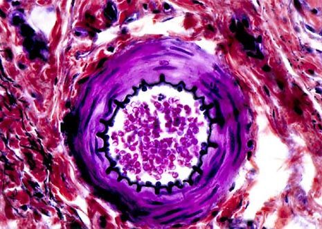
Arteriole, TEM - Stock Image - C015/1018 - Science Photo Library

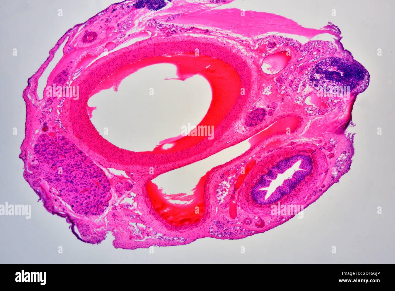
Mammal. Arteriole and venule. Transverse section. 500X - Mammals - Mammals  - Circulatory system - Other systems - Comparative anatomy of Vertebrates -  Animal histology - Photos
Mammal. Arteriole and venule. Transverse section. 500X - Mammals - Mammals - Circulatory system - Other systems - Comparative anatomy of Vertebrates - Animal histology - Photos
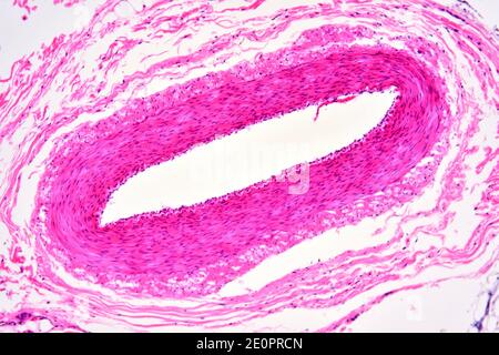
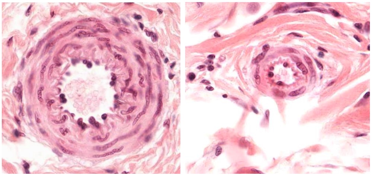
File:"Onion-skin" renal arteriole.jpg - Wikipedia

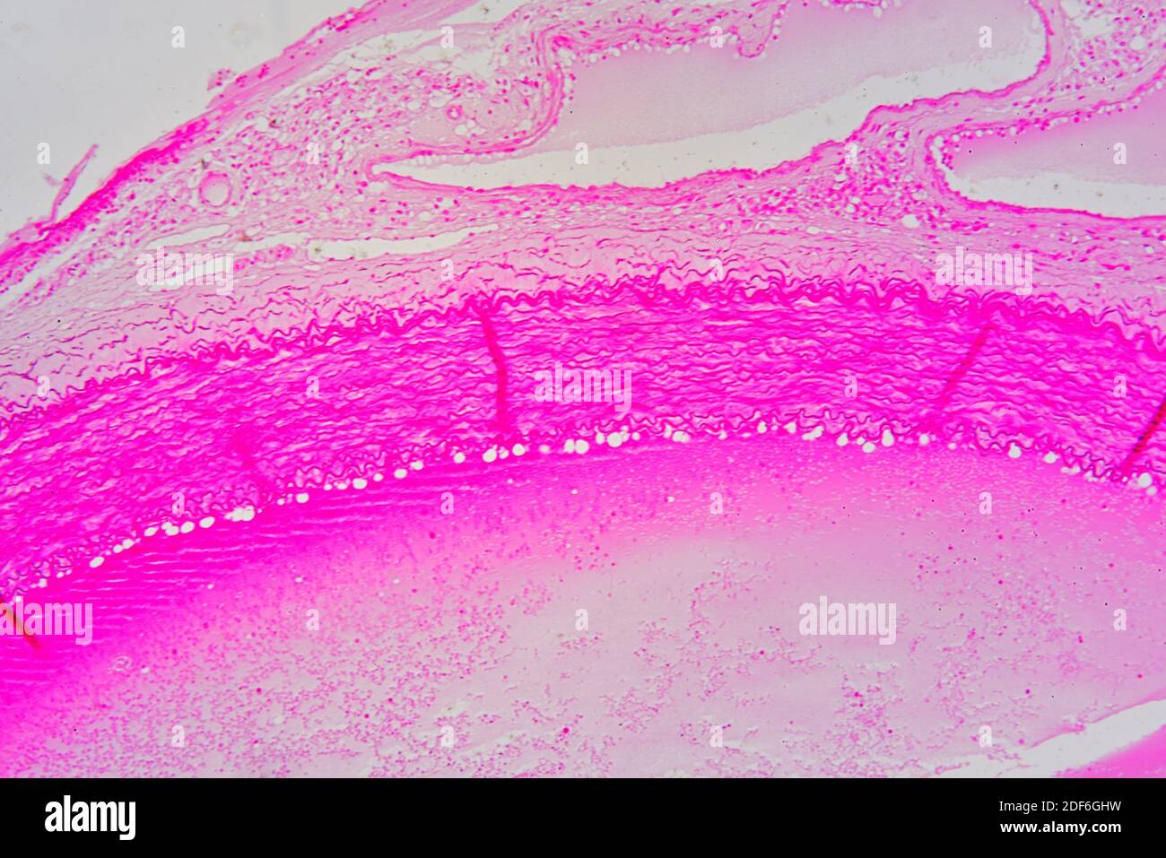
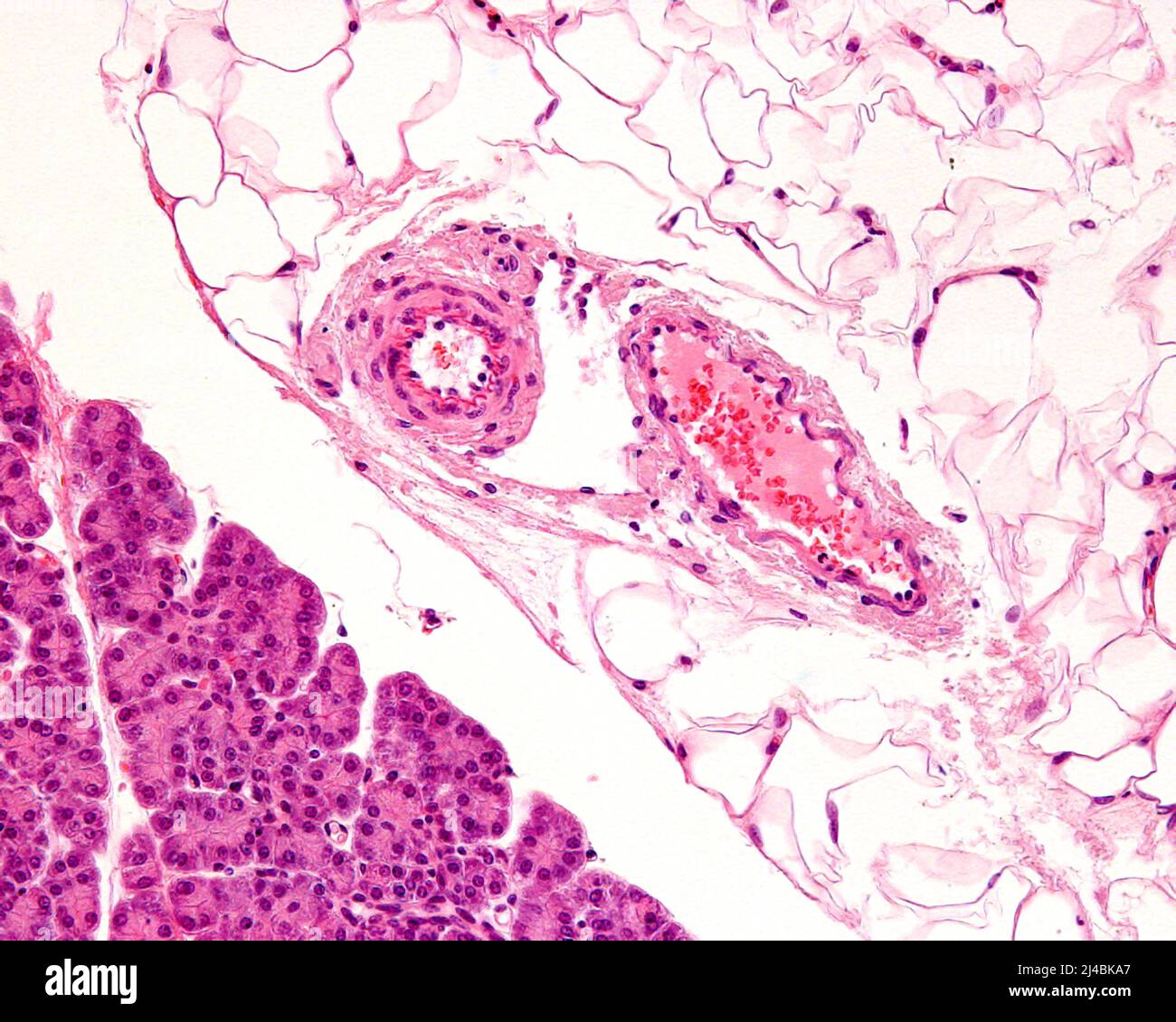
Light microscope micrograph showing the tunica albuginea of a human  testicle. Below the external fibrous layer is the tunica vasculosa and the  seminif Stock Photo - Alamy
Light microscope micrograph showing the tunica albuginea of a human testicle. Below the external fibrous layer is the tunica vasculosa and the seminif Stock Photo - Alamy
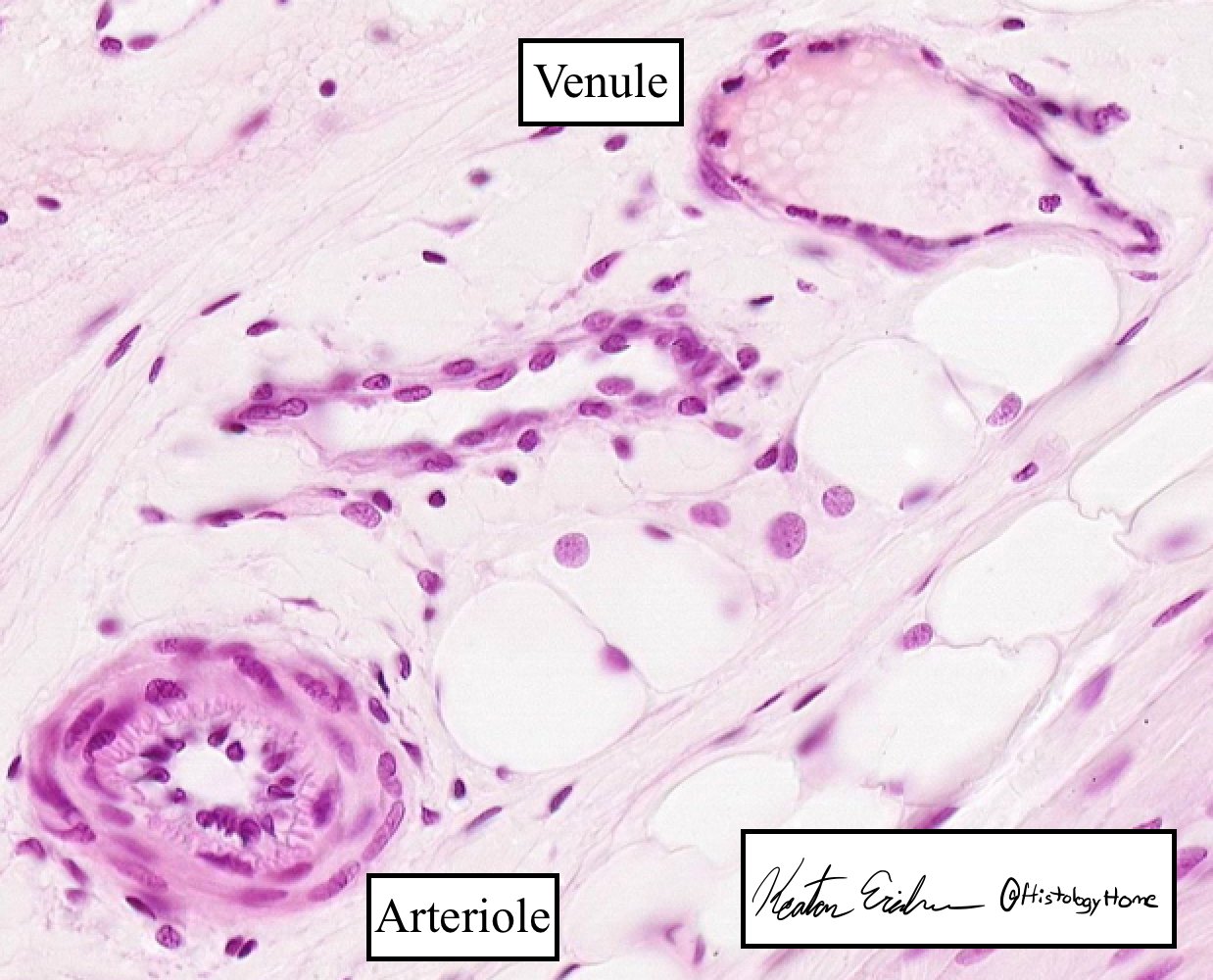
Arteriole Stained Thin Section | Olympus LS

Arteriols Dr.Jastrow's electron microscopic atlas

Human Structure Virtual Microscopy

Photomicrograph artery and vein hi-res stock photography and images - Alamy

Animal organs. Cardiovascular system. Arteriole. Atlas of plant and animal  histology.
Animal organs. Cardiovascular system. Arteriole. Atlas of plant and animal histology.

Arteriole, TEM - Stock Image - C043/5439 - Science Photo Library

Microscopio virtual histoemb - Aparato Cardiovascular

Image of an arteriole (A), venule (V), and lymph vessel (L) in the... |  Download Scientific Diagram
Image of an arteriole (A), venule (V), and lymph vessel (L) in the... | Download Scientific Diagram

Histology Headquarters on X: "Venules are part of the microvasculature, the  first branches after the capillary bed. These vessels have extremely thin  walls! Almost no tunica media is present. Note the contrasting
Histology Headquarters on X: "Venules are part of the microvasculature, the first branches after the capillary bed. These vessels have extremely thin walls! Almost no tunica media is present. Note the contrasting

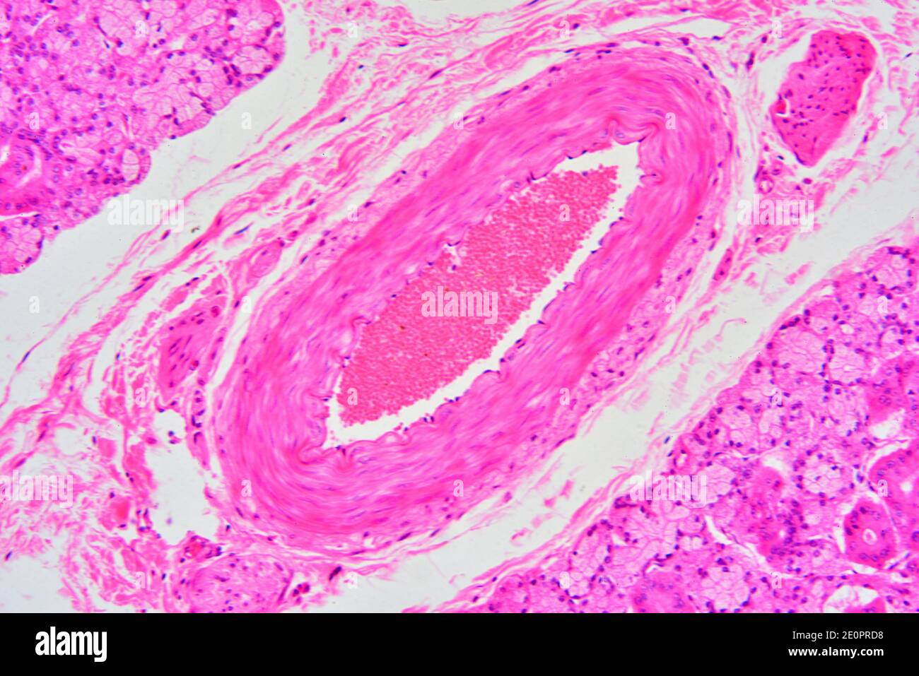
Human arteriole from tongue surrounded by secretory glands. X125 at 10 cm  wide Stock Photo - Alamy
Human arteriole from tongue surrounded by secretory glands. X125 at 10 cm wide Stock Photo - Alamy

Arteriole (blood vessel) wall detail. Optical microscope X100 Stock Photo -  Alamy
Arteriole (blood vessel) wall detail. Optical microscope X100 Stock Photo - Alamy

Microscopio virtual histoemb - Aparato Cardiovascular

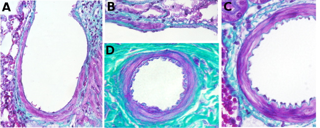
Arteries: Histology | Concise Medical Knowledge

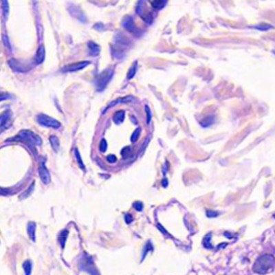
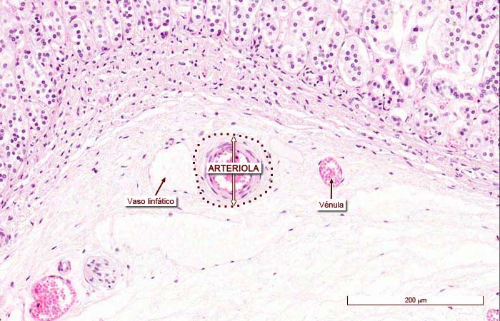
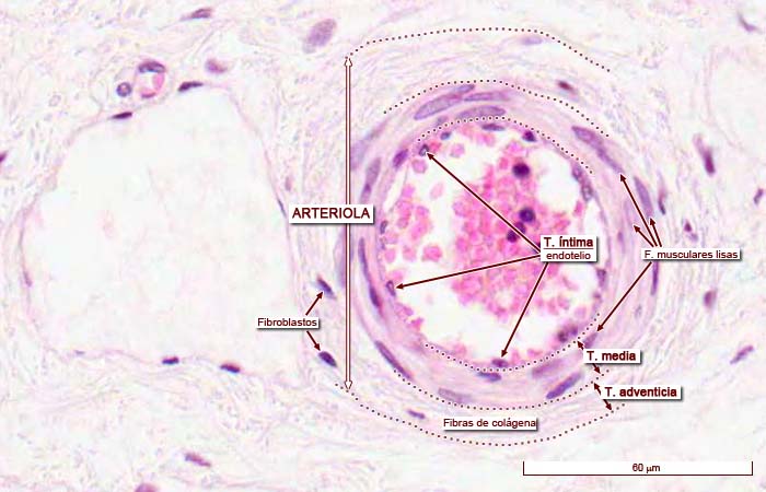
Arteriola
Arteriola

Human Structure Virtual Microscopy

Órganos animales. Cardiovascular: arteriola. Atlas de Histología Vegetal y  Animal
Órganos animales. Cardiovascular: arteriola. Atlas de Histología Vegetal y Animal

Arteriole and venule, light micrograph Stock Photo - Alamy

Microscopio virtual histoemb - Aparato Cardiovascular

Man. Salivary gland. Parotid. Transverse section. 250X - Salivary gland -  Man - Mammals - Digestive system - Other systems - Comparative anatomy of  Vertebrates - Animal histology - Photos
Man. Salivary gland. Parotid. Transverse section. 250X - Salivary gland - Man - Mammals - Digestive system - Other systems - Comparative anatomy of Vertebrates - Animal histology - Photos

Arteriola
Arteriola